Tiếng Việt

While both Cone Beam Computed Tomography (CBCT) and traditional X-rays serve as vital diagnostic tools, the depth of clinical insight they provide differs. Discover how CBCT imaging elevates diagnostic precision – and why it forms the foundation of safe, predictable dental implant surgery at Worldwide Dental Hospital.
When preparing for dental implants or complex procedures, patients are frequently advised to undergo a 3D Cone Beam CT scan. This often prompts two common questions:
“I’ve already had an X-ray, why do I still need a CT scan?”
“What makes these two imaging techniques different?”
While both X-rays and Cone Beam CT are imaging tools, they yield fundamentally different levels of clinical data. For simple treatment cases, standard 2D radiographs offer adequate diagnostic value. However, for advanced procedures requiring high precision – such as full-arch implant placement, complex wisdom tooth extractions, or comprehensive orthodontics – 3D CBCT imaging is the non-negotiable gold standard.
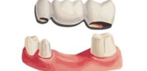
COMPARING X-RAY AND CBCT IN DENTAL DIAGNOSTICS
While both methods share the fundamental principle of using X-rays to capture dental and jawbone structures, they differ significantly in the depth of diagnostic information available to the clinician.
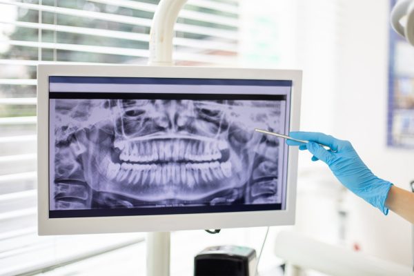
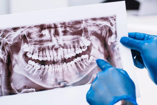
Standard X-rays primarily yield two-dimensional (2D) images, whereas CBCT reconstructs the entire maxillofacial region in three dimensions (3D). This enables clinicians to evaluate anatomical structures across multiple dimensions, rather than being restricted to a single plane.
DIAGNOSTIC FINDINGS PROVIDED BY X-RAYS
X-ray is a widely utilized modality in dentistry that continues to play a critical diagnostic role.
Through 2D radiographs, clinicians can:
- Evaluate dental caries
- Observe root structures
- Detect periapical inflammation
- Monitor erupting teeth
- Assess overall tooth count
For routine dental interventions – such as fillings, endodontic therapy, or simple extractions – standard X-ray scans typically provide sufficient diagnostic value.
However, as two-dimensional representations, spatial overlap of anatomical structures is inherent, restricting their diagnostic capabilities in complex clinical cases.
THE CBCT ADVANTAGE: HIGH-PRECISION 3D ANATOMICAL MAPPING
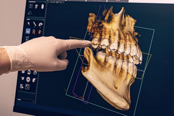
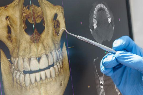
CBCT technology utilizes a cone-shaped X-ray beam to capture hundreds of cross-sectional images in a single rotation, digitally reconstructing a complete 3D model of the patient’s maxillofacial area.
Consequently, clinicians can evaluate:
- Bone height
- Bone width
- Bone density
- Nerve pathways
- Maxillary sinus location
- Spatial relationships between anatomical structures
Rather than merely revealing anatomical presence, CBCT delivers exact spatial intelligence – identifying precise location, depth, and sub-millimeter distances. This high-definition mapping provides a level of predictability that traditional X-rays simply cannot achieve.
THE CRITICAL ROLE OF CBCT IN DENTAL IMPLANT SURGERY
Dental implant placement involves anchoring a titanium post into the jawbone to function as an artificial root for a missing tooth.
Procedure safety relies on precise pre-surgical mapping:
- Remaining bone volume
- Optimal implant dimensions (length and diameter)
- Distance to vital nerve pathways
- Maxillary sinus position
- The necessity for bone grafting or sinus augmentation
These critical diagnostic metrics can only be accurately quantified using CBCT scans. Relying solely on X-ray scans risks misjudging bone width and spatial contour, ultimately compromising the precision of treatment planning.
EXTENDED CLINICAL APPLICATIONS OF CBCT TECHNOLOGY
Beyond implant dentistry, CBCT technology serves as an indispensable diagnostic tool across various specialized disciplines.
Orthodontics
CBCT enables orthodontists to:
- Evaluate root positioning.
- Detect impacted teeth.
- Analyze jaw relationships.
- Track tooth movement.
This facilitates safer, more effective treatment protocols.
Wisdom Tooth Extraction
For malpositioned or impacted wisdom teeth, CBCT accurately visualizes:
- Tooth orientation.
- Proximity to adjacent neural structures.
- Surgical complexity.
This enables clinicians to select appropriate extraction methods and minimize the risk of complications.
Oral Surgery & Restorative Care
CBCT technology provides the foundation for advanced treatment planning, including:
- Bone grafting.
- Sinus lifts.
- Maxillofacial surgeries.
- Surgical guide design.
X-RAY VS. CBCT IMAGING
| Criteria | X-ray | CBCT |
|---|---|---|
| Imaging Dimensionality | 2D | 3D |
| Bone Width Assessment | Not assessable | Assessable |
| Bone Density Evaluation | Limited | Highly accurate |
| Nerve & Sinus Mapping | Limited | Clear |
| Implant Treatment Planning | Basic / Preliminary | Gold standard |
| Clinical Scope | General dentistry | Implants, orthodontic, surgery, & complex cases |
DETERMINING WHEN A CBCT SCAN IS TRULY REQUIRED
CBCT scans are prescribed strictly when clinically necessary, not as a routine.
For instance:
- Routine procedures like fillings or dental cleanings do not require CT scans.
- Root canals or simple extractions typically rely on standard X-rays.
- Complex treatments – such as implant placement, orthodontics, impacted teeth, or oral surgery – demand CBCT imaging to ensure diagnostic precision.
Diagnostic imaging is always guided by actual diagnostic necessity, rather than taking scans indiscriminately.
SAFETY IN CBCT IMAGING
This is a primary concern for many patients.
Modern dental CBCT systems feature a targeted Field of View (FOV) focused specifically on the maxillofacial region. This maximizes diagnostic precision while strictly minimizing unnecessary radiation exposure.
Clinicians prescribe scans only when the diagnostic benefits outweigh potential risks, carefully selecting the appropriate scan scope for each individual case. Patients can therefore feel completely at ease when recommended for this procedure.
INTEGRATING CBCT INTO PERSONALIZED TREATMENT PLANNING AT WORLDWIDE
At Worldwide Dental Hospital, CBCT imaging goes beyond basic visualization – it serves as the cornerstone of precise, upfront treatment planning.
For implant procedures, CBCT datasets are integrated with digital intraoral scans and guided-surgery software to evaluate bone density, map precise implant placement, and select optimal fixture dimensions. When clinically indicated, custom surgical guides are deployed to ensure absolute surgical accuracy.
Combining advanced diagnostic technology with extensive clinical expertise, we tailor every treatment plan strictly to each patient’s unique anatomical profile.
CONCLUSION
X-rays and CBCT are both essential diagnostic tools in modern dentistry, yet each serves a distinct clinical purpose. While 2D X-rays provide a broad baseline overview, CBCT delivers detailed three-dimensional imaging – enabling clinicians to design treatment plans with superior precision, particularly for implants and complex oral surgeries.
If you are considering dental implants, orthodontic therapy, or comprehensive maxillofacial care, visit Worldwide Dental Hospital for a thorough clinical evaluation and personalized diagnostic assessment. Precise diagnostic imaging today is the cornerstone of a safe, predictable, and long-term successful treatment outcome.